ASD Closure Surgery in India
Overview:
ASD (Atrial Septal Defect) Closure Surgery addresses a congenital heart defect where a hole persists in the wall separating the heart’s upper chambers. This defect can lead to abnormal blood flow between the heart and lungs. In India, the cost of ASD closure surgery ranges from USD 4900 to USD 5500. The procedure typically requires ten days in the hospital and another ten days for recovery.
Pre-Surgery Requirements:
Before surgery, patients undergo several tests to evaluate heart function and defect severity. These include a chest X-ray, physical examination, electrocardiogram (ECG), and echocardiogram. These assessments help in planning the most effective treatment approach.
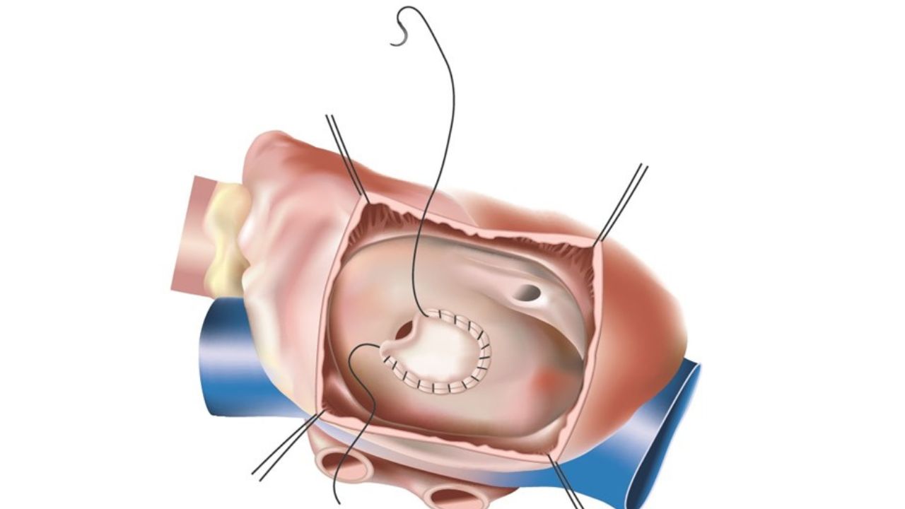
Types of ASD Closure Surgery:
- Open Surgery:
- Performed under general anesthesia with the aid of a heart-lung machine.
- Surgeons make an incision to access the heart, either closing smaller defects with sutures or using a patch for larger ones.
- Minimally Invasive Surgery:
- Conducted through a small chest incision (4-6 cm).
- Uses advanced equipment and an endoscope for visualization.
- Patients experience faster recovery and minimal scarring.
Factors Affecting Cost:
The cost of ASD closure surgery in India can vary depending on the city, hospital, and complexity of the case. In cities like Chennai, costs might differ based on the procedure’s specifics and healthcare facilities.
Treatment Options:
- Medication: While medication cannot correct the defect, it can manage symptoms. Common medications include beta-blockers for heart rate control and anticoagulants to prevent blood clots.
- Surgical Repair:
- Open Surgery: Involves direct access to the heart to close the defect.
- Minimally Invasive Surgery: Less invasive with a quicker recovery time, using a small incision and specialized tools.
Post-Surgery Care:
Post-operative care includes pain management, monitoring for complications, and follow-up visits to ensure proper healing. Patients are advised to avoid strenuous activities to support recovery.
Additional Considerations:
ASD Closure Surgery is highly successful, with a success rate of 98.5%. The choice between open and minimally invasive surgery depends on the defect’s size, location, and patient health. Consulting with a cardiologist or cardiac surgeon is crucial for personalized treatment planning.
In summary, ASD closure surgery in India offers effective treatment for a common congenital heart defect, with options for both traditional and minimally invasive approaches.